Introduction
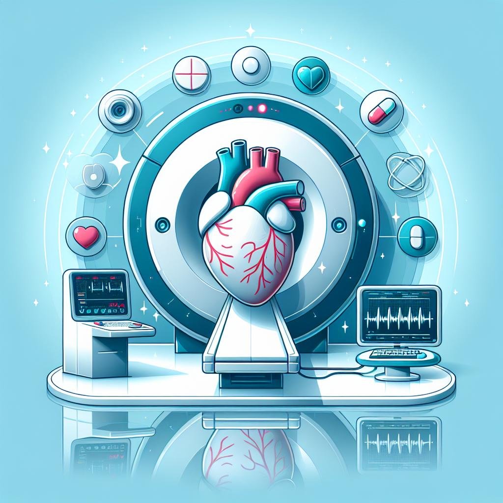
Accurate diagnosis is the foundation of effective cardiac care. Over the past few decades, significant advancements in cardiac imaging technologies have transformed the way healthcare professionals diagnose and manage heart conditions. Three key technologies have become essential tools in the field of cardiology: Computed Tomography (CT) scans, Magnetic Resonance Imaging (MRI), and echocardiography. These imaging methods have greatly improved diagnostic accuracy and led to better patient outcomes.
CT scans use X-rays and computer processing to create detailed cross-sectional images of the heart and blood vessels. This technology allows doctors to see the structure of the heart and identify any abnormalities or blockages in the coronary arteries. CT scans are particularly useful for detecting calcium buildup in the arteries, which can be an early sign of heart disease.
MRI uses powerful magnets and radio waves to produce highly detailed images of the heart and surrounding tissues. This technology is especially valuable for examining the heart’s structure and function, as well as assessing blood flow and detecting scar tissue from previous heart attacks. MRI can provide information about the heart that other imaging methods cannot, making it an important tool for diagnosing complex heart conditions.
Echocardiography uses high-frequency sound waves to create real-time images of the heart. This non-invasive technique allows doctors to see how the heart is beating and pumping blood. Echocardiograms can help identify problems with heart valves, chamber size, and overall heart function. They are often used to diagnose heart murmurs, valve disorders, and other cardiac abnormalities.
These advanced imaging technologies have revolutionized cardiac care by providing more accurate and detailed information about the heart’s structure and function. This improved diagnostic capability allows doctors to detect heart problems earlier and develop more effective treatment plans. As a result, patients can receive more targeted and timely interventions, leading to better overall outcomes.
In the following sections, we will explore the evolution of cardiac imaging, delve deeper into the principles and applications of CT scans, MRI, and echocardiography, and examine how these advancements are shaping the future of cardiac care. By understanding these imaging technologies, we can appreciate their importance in modern cardiology and their potential to improve heart health for countless patients.
The Evolution of Cardiac Imaging
Early Imaging Techniques
Cardiac imaging has undergone remarkable transformations since its early days. The journey began with X-rays and fluoroscopy, which were revolutionary for their time. X-rays allowed doctors to see the heart’s silhouette and detect enlargements or changes in shape. However, they could only provide static images, limiting their usefulness in understanding the heart’s dynamic function. Fluoroscopy, on the other hand, offered real-time imaging of the heart’s movement. This was a significant improvement, but the images lacked detail and clarity. Despite these limitations, these early techniques laid the foundation for future advancements in cardiac imaging.
The introduction of CT scans marked a significant leap forward in cardiac imaging technology. Early CT scanners were slow and produced images with limited resolution. They took several seconds to capture a single slice of the heart, which made it challenging to get clear pictures of this constantly moving organ. However, these early CT scans were crucial in demonstrating the potential of this technology for cardiac imaging. They showed that it was possible to get detailed cross-sectional images of the heart, paving the way for future improvements in speed and resolution.
Advancements in CT Scans
The field of cardiac imaging has been revolutionized by the development of high-speed and multi-detector CT scans. These advanced machines can capture images of the heart in a fraction of a second, effectively “freezing” its motion. This speed allows for clearer, more detailed images that are less affected by the heart’s movement. Multi-detector CT scans use multiple rows of detectors to capture several slices of the heart simultaneously, greatly increasing the amount of information gathered in a single scan.
Cardiac CT angiography (CTA) has emerged as a powerful diagnostic tool for coronary artery disease. This technique involves injecting a contrast dye into the bloodstream and then using CT to create detailed 3D images of the heart and its blood vessels. CTA can detect even small blockages in the coronary arteries, helping doctors identify potential problems before they become serious. The procedure is quick, usually taking only a few minutes, which helps reduce patient anxiety compared to more invasive procedures like cardiac catheterization.
MRI and Echocardiography
Magnetic Resonance Imaging (MRI) has made significant strides in cardiac imaging. MRI uses powerful magnets and radio waves to create detailed images of the heart without using radiation. This makes it a safe option for repeated use or for imaging children and pregnant women. Cardiac MRI is particularly good at showing the structure of the heart muscle, making it useful for diagnosing conditions like cardiomyopathies (diseases of the heart muscle). It can also show blood flow through the heart and major blood vessels, helping doctors assess valve function and detect blood clots.
Echocardiography has also seen major improvements over the years. This technique uses ultrasound waves to create moving pictures of the heart. Modern echocardiography machines can produce high-resolution, real-time images of the heart’s chambers, valves, and blood flow. One of the biggest advantages of echocardiography is its portability. Small, handheld devices can now perform basic echocardiograms at a patient’s bedside or in emergency situations. This technology has made it possible to quickly assess heart function in critical situations, potentially saving lives. Additionally, 3D echocardiography has emerged as a powerful tool for visualizing complex heart structures and planning surgical interventions.
CT Scans in Cardiac Imaging
Principles of CT Scans
Computed Tomography (CT) scans are powerful tools in cardiac imaging that use X-rays to create detailed pictures of the heart and its surrounding structures. The process begins with the patient receiving an injection of a special contrast dye into their arm. This dye helps to make the blood vessels and heart structures more visible in the images. Once the dye is injected, the patient lies down on a table that slides into the CT scanner. The scanner looks like a large donut and has an X-ray tube that rotates around the patient’s body. As the X-ray tube moves, it takes many pictures of thin slices of the heart from different angles. These slices are then combined by a computer to create a three-dimensional view of the heart. This 3D image allows doctors to see even tiny blockages in the coronary arteries, which are the blood vessels that supply oxygen to the heart muscle.
Advantages of CT Scans
CT scans offer many benefits for cardiac imaging. One of the biggest advantages is their speed. High-speed CT scanners can capture images of the heart in just a few minutes, which is much faster than traditional cardiac catheterization procedures. This quick imaging time means less discomfort for patients and allows doctors to see results more quickly. CT scans also provide very detailed pictures of the heart’s anatomy and how well it’s working. This makes them excellent for diagnosing coronary artery disease (CAD) and other heart problems. Another great feature of CT scans is that they can show the heart and coronary arteries from any angle. Doctors can even create three-dimensional views, which helps them plan treatments more accurately. For example, if a patient needs heart surgery, the 3D images can help the surgeon know exactly where to operate.
Limitations and Challenges
While CT scans are very useful, they do have some drawbacks. One of the main concerns is radiation exposure. When a person gets a CT scan, they are exposed to a small amount of radiation from the X-rays. Although modern CT scanners have reduced this risk, doctors still try to limit the number of scans a person gets, especially for children and pregnant women. Another challenge is the use of contrast dye. Some people may have allergic reactions to the dye, and it can be harmful to people with kidney problems. Doctors need to check a patient’s kidney function before giving them the contrast dye. Reading CT scan images also requires special training. Doctors who look at these images need to study for many years to understand what they’re seeing. If an image is not read correctly, there’s a chance of making a wrong diagnosis. This is why it’s important for experienced specialists to review CT scan results. Despite these challenges, CT scans remain a valuable tool in cardiac imaging when used appropriately.
MRI in Cardiac Imaging
Principles of MRI
Magnetic Resonance Imaging (MRI) is a powerful tool in cardiac imaging that uses strong magnetic fields and radio waves to create detailed pictures of the heart. Unlike CT scans, MRI does not use ionizing radiation, making it a safer option for patients who need repeated imaging over time. During an MRI scan, the patient lies on a comfortable table that slides into a large, tube-shaped machine. The machine generates a strong magnetic field around the patient, causing the hydrogen atoms in their body to align. Radio waves are then sent through the body, causing these atoms to produce faint signals. These signals are detected by the MRI machine and turned into detailed images by a computer.
The MRI procedure can sometimes cause anxiety in patients who feel uncomfortable in enclosed spaces. To address this issue, some hospitals now offer open MRI scanners, which have a more spacious design. However, these open scanners are not typically used for heart imaging because they may not provide the same level of detail as traditional MRI machines.
Advantages of MRI
MRI offers several important benefits in cardiac imaging. First, it provides very clear, high-resolution pictures of the heart’s structure and how it’s working. This makes MRI the best choice for many types of heart tests. Doctors can use MRI to see things like heart valve problems, unusual heart shapes, and blood clots in the heart with great accuracy.
Another big advantage of MRI is that it doesn’t use harmful radiation. This is especially helpful for patients who need many heart scans over a long time. For example, people with chronic heart conditions can have regular MRI scans without worrying about the risks of repeated radiation exposure.
MRI can also show how blood flows through the heart and blood vessels. This helps doctors understand if there are any blockages or problems with blood flow. Additionally, MRI can measure how well the heart is pumping blood, which is important for diagnosing and monitoring heart failure.
Limitations and Challenges
While MRI is a very useful tool, it does have some drawbacks. Many people feel scared or uncomfortable in the small space of the MRI machine. This fear, called claustrophobia, can be so strong that some patients need medicine to help them relax during the scan.
Another challenge is that MRI doesn’t work well with metal. Things like jewelry, dental fillings, and some tattoo inks can affect the quality of the images. More seriously, people with certain medical devices like pacemakers or metal implants usually can’t have MRI scans because the strong magnets could cause problems.
MRI scans also take a long time, often 30 to 60 minutes or more. This can be hard for people who have trouble lying still for long periods. The machines are also very noisy, which some patients find unpleasant even with earplugs.
Lastly, reading MRI scans of the heart requires special training. Not all doctors can do this, which means that hospitals need specially trained staff to interpret the results. This can sometimes lead to delays in getting test results back to patients.
Echocardiography in Cardiac Imaging
Principles of Echocardiography
Echocardiography is a powerful cardiac imaging technique that uses ultrasound waves to create detailed pictures of the heart. This method involves placing a small device called a transducer on the patient’s chest. The transducer sends out high-frequency sound waves that bounce off the heart’s structures and return to the device. These echoes are then converted into moving images on a screen, allowing doctors to see the heart in action.
One of the key advantages of echocardiography is its versatility. It can be performed in various settings, from hospital rooms to emergency departments, making it an essential tool for quick diagnoses. The technique is particularly useful in emergency situations where rapid assessment of heart function is crucial.
Doppler echocardiography is a specialized form of this imaging method. It measures the speed and direction of blood flow through the heart and blood vessels. This additional information helps doctors evaluate how well the heart is pumping and whether there are any issues with blood flow, such as leaky valves or blockages.
Advantages of Echocardiography
Echocardiography offers numerous benefits in cardiac imaging. First and foremost, it is a very safe procedure. Unlike some other imaging techniques, it doesn’t use radiation, making it suitable for repeated use and safe for pregnant women. The equipment is also portable, allowing doctors to perform the test at a patient’s bedside if necessary.
Another significant advantage is the real-time nature of the images. Doctors can see the heart beating and blood flowing through its chambers as it happens. This live view is invaluable for diagnosing conditions that may only be apparent when the heart is in motion.
Echocardiography is also relatively inexpensive compared to other cardiac imaging methods. This makes it more accessible to a wider range of patients and healthcare facilities. The procedure is non-invasive, meaning it doesn’t require any incisions or injections, which adds to patient comfort and reduces risks.
The technique is particularly good at detecting certain heart problems. It can show the size and shape of the heart, how well the heart’s chambers and valves are working, and whether there are any abnormal growths or fluid around the heart. Echocardiography can also identify issues such as:
- Heart valve problems, including narrowing or leaking of valves
- Congenital heart defects
- Weakening or thickening of heart muscle
- Blood clots inside the heart
- Fluid buildup in the sac around the heart (pericardial effusion)
- Infections affecting the heart valves (endocarditis)
Limitations and Challenges
While echocardiography is a valuable tool, it does have some limitations. The quality of the images can vary depending on several factors. The skill and experience of the person performing the test (known as a sonographer) play a crucial role. An experienced sonographer can often obtain clearer, more detailed images than a less experienced one.
Patient factors can also affect image quality. For example, obesity, lung disease, or chest wall deformities can make it harder to get clear pictures of the heart. In some cases, these issues might require the use of alternative imaging methods.
Another limitation is the level of detail provided by echocardiography. While it offers excellent images of the heart’s structure and function, it may not provide as much detail as other imaging techniques like CT scans or MRI for certain conditions. For instance, it might not be as effective at detecting small blood clots or minor changes in the coronary arteries.
Interpretation of echocardiograms requires specialized training and experience. Misinterpretation of images can lead to incorrect diagnoses, which is why it’s crucial for echocardiograms to be read by qualified cardiologists or specially trained radiologists. This need for expertise can sometimes lead to delays in diagnosis, especially in smaller healthcare facilities that might not have specialists available around the clock.
Despite these challenges, echocardiography remains a cornerstone of cardiac imaging due to its safety, accessibility, and ability to provide real-time information about heart function. Ongoing advancements in technology continue to improve image quality and expand the capabilities of this important diagnostic tool.
Improving Diagnosis with Advanced Imaging
Enhanced Visualization
Advanced cardiac imaging techniques have revolutionized the way doctors see and understand the heart. These methods, including CT scans, MRI, and echocardiography, provide incredibly detailed pictures of the heart’s structure and how it works. CT scans use X-rays to create cross-sectional images of the heart, showing its shape and any problems with blood vessels. MRI uses powerful magnets and radio waves to produce clear, detailed images of the heart’s soft tissues. Echocardiography uses sound waves to create moving pictures of the heart in action, allowing doctors to see how it beats and pumps blood in real-time.
These improved imaging techniques help doctors spot even tiny issues that might have been missed before. For example, they can see small blockages in the arteries that supply blood to the heart, problems with heart valves, or unusual heart rhythms. This enhanced visualization means doctors can find and treat heart problems earlier, which often leads to better results for patients.
Accurate Diagnosis
Getting the right diagnosis is super important when it comes to heart problems. The new imaging methods we have now make it much easier for doctors to figure out exactly what’s wrong. CT scans are really good at finding small blockages in the arteries around the heart. This helps doctors know if a patient needs treatment to open up those arteries. MRI scans are great for looking at the heart muscle and valves. They can show if parts of the heart aren’t working right or if there are problems with how the heart formed.
Echocardiography is special because it lets doctors see the heart moving in real-time. This is really helpful for finding problems that only show up when the heart is beating, like some valve issues or problems with how the heart walls move. All of these tools working together mean that doctors can be much more sure about what’s causing a patient’s heart problems. This leads to better treatment plans and helps patients get the right care faster.
Personalized Medicine
Advanced imaging is helping doctors create treatment plans that are just right for each patient. This is called personalized medicine, and it’s a big deal in heart care. By getting such detailed pictures of a person’s heart, doctors can figure out the best way to help that specific patient. For instance, CT scans can show which patients would benefit most from a procedure called angioplasty, where doctors open up blocked arteries. MRI scans help doctors decide the best spots to put devices like pacemakers or defibrillators, which help control heart rhythms.
Echocardiography is really useful for checking how well treatments are working. Doctors can use it to see if a patient’s heart is getting stronger after starting a new medicine or having a procedure. If things aren’t improving as much as they should, the doctor can quickly change the treatment plan. This ability to tailor treatments and keep a close eye on how they’re working means patients have a better chance of getting healthy and staying that way.
Future Directions in Cardiac Imaging
Emerging Technologies
The field of cardiac imaging is on the brink of a revolution, thanks to emerging technologies that promise to enhance diagnosis and treatment of heart conditions. Artificial intelligence (AI) and machine learning (ML) are at the forefront of these advancements, offering new ways to process and interpret medical images. These smart technologies can quickly analyze large amounts of data, spotting patterns and details that human eyes might miss. This can lead to faster and more accurate diagnoses, giving doctors better tools to help their patients.
Another exciting development is hybrid imaging. This technique combines different types of scans, like PET (Positron Emission Tomography) and CT (Computed Tomography), to create a more complete picture of the heart. By merging these images, doctors can see both the structure and function of the heart at the same time. This gives them a better understanding of what’s happening inside the body, which can lead to more precise treatments.
3D printing is also making waves in cardiac imaging. Doctors can now create detailed, three-dimensional models of a patient’s heart using imaging data. These models help surgeons plan complex procedures and can be used to explain conditions to patients in a way that’s easy to understand.
Potential Applications
The new technologies in cardiac imaging have many useful applications. AI and ML can help doctors spot heart problems earlier, even before symptoms appear. This early detection can lead to better treatment outcomes and potentially save lives. These smart systems can also help predict which patients are at higher risk for certain heart conditions, allowing for preventive care.
Hybrid imaging techniques are particularly useful for complex cases. For example, they can help doctors see exactly where a problem is in the heart and how it’s affecting blood flow. This detailed information can guide treatment decisions and make surgeries safer and more effective.
3D printed heart models have several exciting uses. Surgeons can practice complicated procedures on these models before operating on real patients. This can make surgeries safer and reduce the time patients spend under anesthesia. These models are also great teaching tools for medical students and can help patients better understand their conditions.
Challenges and Opportunities
While these new technologies offer many benefits, they also come with challenges. One big hurdle is the need for large amounts of data to train AI and ML systems. Gathering this data while protecting patient privacy is a complex task. There’s also a need for standards in how this data is collected and used to ensure that the AI systems are accurate and fair for all patients.
Another challenge is the cost of new equipment and training. Hybrid imaging machines and 3D printers can be expensive, and not all hospitals may be able to afford them. Doctors and technicians also need special training to use these new tools effectively.
Despite these challenges, the opportunities are enormous. As these technologies become more common, they could lead to more personalized care for heart patients. Doctors could use detailed imaging data to create treatment plans tailored to each person’s unique heart structure and condition.
There’s also potential for these technologies to make cardiac care more accessible. For example, AI could help bring expert-level image analysis to smaller hospitals or remote areas that don’t have specialist doctors.
As research continues, we may see even more exciting developments. Scientists are working on ways to use imaging to detect heart problems at the molecular level, potentially catching issues before they cause any damage. This could open up new possibilities for preventing heart disease and improving overall heart health.
Conclusion
Advancements in cardiac imaging technologies have greatly improved how doctors diagnose heart problems and help patients. CT scans, MRI, and echocardiography have become very important tools for heart doctors. These technologies allow doctors to see inside the heart more clearly, which helps them make more accurate diagnoses. They also help doctors create personalized treatment plans for each patient.
CT scans use X-rays to create detailed pictures of the heart. This helps doctors see things like blocked arteries or problems with heart valves. MRI uses strong magnets to make clear images of the heart’s structure and how it’s working. Echocardiography uses sound waves to create moving pictures of the heart, showing how it beats and pumps blood.
New technologies are also making cardiac imaging even better. Artificial intelligence (AI) and machine learning (ML) are helping doctors analyze images faster and more accurately. These computer programs can spot tiny details that humans might miss. Hybrid imaging combines different types of scans to give doctors even more information about a patient’s heart.
As these technologies continue to improve, they will help doctors find heart problems earlier and treat them more effectively. This means patients can get better care and have a better chance of staying healthy. Cardiac imaging is always changing and getting better, which is great news for anyone with heart issues.
Doctors and scientists are always working on new ways to see inside the heart. They want to make the tests faster, more comfortable for patients, and even more accurate. As they keep improving these imaging tools, it will be easier to spot heart problems early and treat them before they become serious.
References
- https://insightsimaging.springeropen.com/articles/10.1007/s13244-010-0049-0
- https://www.webmd.com/heart/features/imaging-heart-new-frontier
- https://www.ncbi.nlm.nih.gov/pmc/articles/PMC9872125/
- https://www.ncbi.nlm.nih.gov/books/NBK448128/
- https://my.clevelandclinic.org/health/diagnostics/16836-cardiac-imaging