Introduction
Why Cardiac Imaging Matters
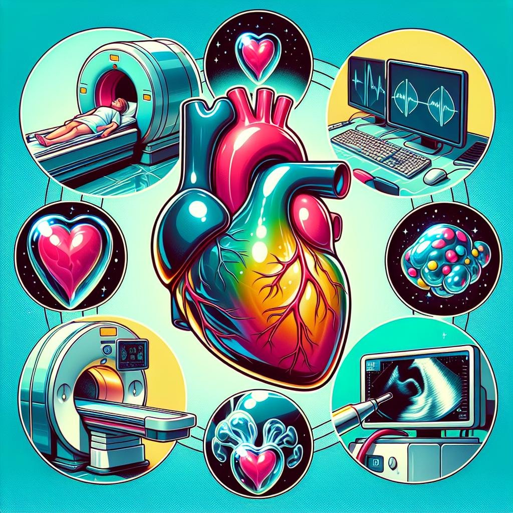
Cardiac imaging is an essential tool that plays a crucial role in diagnosing and managing various heart conditions. These advanced techniques provide healthcare providers with detailed and clear pictures of the heart and its surrounding structures. By using cardiac imaging, doctors can accurately diagnose heart problems and create effective treatment plans tailored to each patient’s needs.
There are several reasons why cardiac imaging is so important:
-
Accurate diagnosis: Cardiac imaging helps doctors identify a wide range of heart conditions. For example, it can detect coronary artery disease, which occurs when the blood vessels supplying the heart become narrowed or blocked. It can also reveal heart failure, where the heart doesn’t pump blood as well as it should. Additionally, cardiac imaging can show structural abnormalities in the heart, such as defects in the heart’s valves or chambers.
-
Monitoring disease progression: For patients with existing heart conditions, cardiac imaging is vital in tracking how their disease changes over time. Regular imaging allows doctors to see if a condition is getting worse or if it’s responding well to treatment. This information helps healthcare providers adjust treatment plans as needed to ensure the best possible outcomes for their patients.
-
Evaluating treatment effectiveness: After starting a new medication or undergoing a procedure, cardiac imaging can show how well the treatment is working. For instance, if a patient has had a stent placed in a blocked artery, follow-up imaging can confirm that blood is flowing properly through the stent.
-
Guiding procedures: Some cardiac imaging techniques can be used during heart procedures to help doctors navigate and make real-time decisions. This improves the accuracy and safety of many heart treatments.
-
Preventive care: In some cases, cardiac imaging can detect heart problems before a person experiences any symptoms. This early detection allows for prompt treatment, potentially preventing more serious heart issues from developing.
By understanding the different types of cardiac imaging techniques and what to expect during these procedures, patients can feel more prepared and less anxious. This knowledge also helps patients make informed decisions about their heart health and actively participate in discussions with their healthcare providers. Cardiac imaging is a powerful tool that continues to advance, offering new ways to look inside the heart and improve patient care.
Types of Cardiac Imaging Techniques
Magnetic Resonance Imaging (MRI)
How MRI Works
Magnetic Resonance Imaging (MRI) is a powerful tool for looking inside the heart. It uses a big magnet and special radio waves to make detailed pictures. During an MRI, you lie on a table that slides into a large, tube-shaped machine. The magnet and radio waves work together to line up tiny parts of your body called atoms. These atoms then send out signals that help create clear images of your heart.
Advantages and Limitations
MRI has many good points. It gives very clear pictures and can show soft parts of the heart really well. It also doesn’t use harmful radiation. However, MRI has some drawbacks too. It can be expensive and isn’t good for people with certain metal items in their body. Some people might feel scared in the small space of the MRI machine. The scan can also take a long time, sometimes up to an hour, and the machine can be very noisy.
Common Uses in Cardiac Diagnosis
Doctors use MRI for many heart tests. It can show how big the heart is and how thick its walls are. MRI can also show how well the heart is pumping blood and if the heart valves are working right. It’s very good at finding problems like weak heart muscles or blocked arteries. Doctors also use MRI to see how the heart reacts to exercise and to check if treatments are working.
Computed Tomography (CT) Scans
How CT Scans Work
CT scans use X-rays and computers to make detailed pictures of the heart. When you get a CT scan, you lie on a table that moves through a big, donut-shaped machine. This machine spins around you, taking many X-ray pictures from different angles. A computer then puts all these pictures together to make a clear image of your heart.
Advantages and Limitations
CT scans have several good points. They are fast and give very clear pictures. They can show both soft parts and hard parts of the heart. Sometimes, doctors use a special dye to make the pictures even clearer. However, CT scans do have some drawbacks. They use X-rays, which can be harmful if you have too many. Also, some people might be allergic to the special dye, and it can cause problems for people with kidney issues.
Common Uses in Cardiac Diagnosis
Doctors often use CT scans to look for blocked arteries in the heart. They can see if there’s any buildup in the arteries that might cause problems. CT scans also show the size and shape of the heart chambers and how well the heart walls are moving. Like MRI, CT scans can help doctors see how the heart reacts to exercise and if treatments are working well.
Echocardiography
How Echocardiography Works
Echocardiography uses sound waves to make pictures of your heart. It’s like the ultrasound used to see babies before they’re born. During an echocardiogram, a doctor or technician puts a special gel on your chest. Then they use a tool called a probe to send sound waves into your chest. These waves bounce off your heart and create moving pictures on a computer screen.
Advantages and Limitations
Echocardiography has many good points. It doesn’t hurt, doesn’t use harmful radiation, and shows your heart moving in real-time. It’s also quick and can be done in a doctor’s office. However, it’s not perfect. The pictures might not be as clear as those from MRI or CT scans. Also, it can be harder to get good pictures if you have a large body or lung problems.
Common Uses in Cardiac Diagnosis
Doctors use echocardiography for many heart tests. It can show if your heart valves are working right or if there are any holes in your heart. It also measures how well your heart is pumping blood. Doctors use it to watch how heart problems change over time and to see if treatments are helping.
Positron Emission Tomography (PET) Scans
How PET Scans Work
PET scans use a special type of energy to make pictures of your heart. Before the scan, you get an injection of a safe, slightly radioactive liquid. This liquid goes to parts of your heart that aren’t getting enough blood. A special camera can see this liquid and make pictures that show where your heart might have problems.
Advantages and Limitations
PET scans are very good at finding areas of your heart that aren’t getting enough blood. They can show problems that other tests might miss. However, PET scans do have some drawbacks. They use a small amount of radiation, which can be harmful if you have too many scans. They can also be expensive and aren’t available at all hospitals.
Common Uses in Cardiac Diagnosis
Doctors often use PET scans to look for blocked arteries in the heart. They can show which parts of the heart aren’t getting enough blood. PET scans also help doctors see how well the heart is working during exercise. They can show if treatments are helping to improve blood flow to the heart.
Other Techniques
There are other ways doctors can look at your heart. One is called angiography. In this test, doctors put a special dye into your heart arteries and take X-ray pictures. Another test is cardiac catheterization. In this test, doctors put a thin tube into your heart arteries to look for blockages. They can also use this tube to fix some heart problems. These tests are more invasive, which means they involve going inside your body. Doctors usually only use them when other tests don’t give enough information or when they need to fix a problem right away.
Preparing for a Cardiac Imaging Procedure
What to Wear
When getting ready for a cardiac imaging procedure, it’s important to choose your clothes carefully. Wear comfortable, loose-fitting clothing that allows easy access to your chest area. This helps the medical team perform the imaging without any obstacles. Avoid wearing jewelry, as it can interfere with the imaging equipment. Metal fasteners on clothing, such as zippers or buttons, can also cause problems during the imaging process. Instead, opt for soft, stretchy fabrics like cotton t-shirts or sweatshirts. Some imaging centers may provide hospital gowns for you to change into before the procedure. If you’re unsure about what to wear, don’t hesitate to ask your healthcare provider for guidance.
Fasting and Medication
Depending on the type of cardiac imaging you’re having, you might need to fast for a certain period before the procedure. Fasting means not eating or drinking anything except water. For example, if you’re having a CT scan, you may need to avoid food and drinks for several hours beforehand. This helps ensure that the images are clear and accurate. It’s crucial to follow your doctor’s instructions about fasting carefully.
Regarding medications, it’s essential to tell your healthcare provider about all the medicines you’re taking. This includes prescription drugs, over-the-counter medications, and even supplements or herbal remedies. Some medications might need to be adjusted or temporarily stopped before the imaging procedure. For instance, if you’re taking diabetes medication, your doctor might give you special instructions on how to manage your blood sugar levels while fasting. Never stop or change your medications without talking to your doctor first.
What to Expect During the Procedure
During a cardiac imaging procedure, you’ll typically lie down on a special table. This table is designed to slide into the imaging machine. For example, if you’re having an MRI (Magnetic Resonance Imaging), you’ll enter a large, tube-shaped machine. The table moves slowly into the machine, positioning your body correctly for the imaging.
Before the procedure starts, a technician will explain everything to you. They’ll tell you how long the imaging will take and what you need to do. Don’t be afraid to ask questions if you’re unsure about anything. The technician wants you to feel comfortable and understand what’s happening.
One of the most important things during the imaging is to stay very still. Moving can blur the images, making them less useful for diagnosis. The technician might ask you to hold your breath for short periods during the imaging. This helps keep your chest area steady for clear pictures.
Some imaging procedures, like ultrasounds, might require a special gel on your skin. This gel helps the ultrasound waves move between the machine and your body. It might feel a bit cold but doesn’t hurt at all.
Remember, the imaging machine might make loud noises. This is normal and nothing to worry about. Some places offer earplugs or headphones to make you more comfortable during the procedure. The most important thing is to relax and follow the technician’s instructions. This will help ensure that the cardiac imaging gives your doctors the best possible information about your heart.
What to Expect During the Diagnosis
Understanding the Results
After completing the cardiac imaging procedure, patients can expect to receive their results from their healthcare provider. These results typically include detailed images of the heart and surrounding structures, which provide valuable information about the organ’s health and function. The healthcare provider will carefully review these images to identify any abnormalities or conditions that may be present. During the follow-up appointment, the provider will explain the results in a clear and understandable manner, using simple language to ensure the patient grasps the information. Patients are encouraged to ask questions about their results to gain a better understanding of their heart health.
Common Findings
Cardiac imaging procedures often reveal various conditions and abnormalities related to heart health. Some of the most common findings include:
-
Coronary artery disease: This occurs when the blood vessels supplying the heart become narrowed or blocked, reducing blood flow to the heart muscle.
-
Heart failure: This condition develops when the heart is unable to pump blood effectively to meet the body’s needs.
-
Valve problems: Issues with the heart’s valves can affect blood flow through the heart chambers.
-
Heart defects: These are structural abnormalities present from birth that can impact heart function.
-
Enlarged heart: An increase in heart size can indicate various underlying conditions.
-
Blood clots: Imaging may reveal the presence of blood clots in the heart or nearby blood vessels.
The healthcare provider will thoroughly explain any findings to the patient, discussing their implications for overall heart health and potential treatment options.
Next Steps
After reviewing the results of the cardiac imaging procedure, the healthcare provider will work with the patient to determine the most appropriate next steps. These may include:
-
Treatment options: Depending on the findings, the provider may recommend various treatments such as medication, lifestyle changes, or medical interventions like angioplasty or stenting.
-
Follow-up care: The healthcare provider will outline a plan for ongoing monitoring and care to ensure the patient’s heart health is properly managed.
-
Additional testing: In some cases, further tests may be necessary to gather more information or confirm a diagnosis.
-
Referral to specialists: If needed, the provider may refer the patient to a cardiologist or other heart specialist for more specialized care.
-
Lifestyle modifications: The healthcare provider may suggest changes to diet, exercise habits, or stress management techniques to improve heart health.
-
Medication adjustments: Based on the imaging results, the provider may recommend starting, stopping, or modifying medications to better manage heart health.
Throughout this process, the healthcare provider will encourage open communication and address any concerns or questions the patient may have about their heart health and treatment plan.
Conclusion
Cardiac imaging techniques play a crucial role in helping doctors understand and treat heart problems. These methods give detailed pictures of the heart and nearby areas, which helps doctors make the right diagnosis and create effective treatment plans. There are several different types of cardiac imaging, each with its own strengths and uses.
When patients know what to expect during these tests, they can feel more comfortable and prepared. It’s important for patients to remember that these imaging procedures are generally safe and painless. Most of the time, patients can go back to their normal activities right after the test is done.
Doctors use cardiac imaging to look for many different heart issues. They can see if there are problems with how the heart is shaped, how it’s working, or if there are any blockages in the blood vessels. This information helps them decide the best way to treat each patient’s unique situation.
Patients should always follow their doctor’s instructions before and after any imaging test. This might include things like not eating for a certain time before the test or avoiding certain medications. It’s also a good idea for patients to write down any questions they have and ask their doctor or the imaging technician. This way, they can fully understand what’s happening and why it’s important for their health.
Remember, cardiac imaging is just one part of taking care of your heart health. It works together with other things like regular check-ups, a healthy diet, exercise, and taking prescribed medications. By using these imaging tools and following their doctor’s advice, patients can take an active role in keeping their hearts healthy and strong.
References
- Heart & Vascular Care. Cardiac Imaging, Testing & Diagnostics. [Accessed 13 Aug 2024].
- Mayo Clinic. Heart disease – Diagnosis and treatment. [Accessed 13 Aug 2024].
- StatPearls. Cardiac Imaging. [Accessed 13 Aug 2024].
- NCBI. Recent technologies in cardiac imaging. [Accessed 13 Aug 2024].
- Cleveland Clinic. Cardiac Imaging: Types, Uses and Procedure Details. [Accessed 13 Aug 2024].
