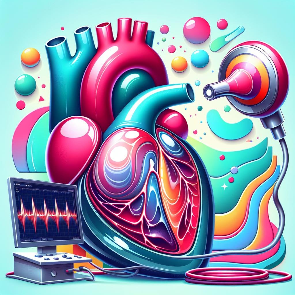
Introduction
Doppler echocardiography is a remarkable medical technology that helps doctors examine the heart without causing any discomfort to patients. This method uses sound waves to create detailed images of the heart and its blood flow. It’s like having a special window that allows doctors to see inside the heart and understand how it’s working.
The technique gets its name from the Doppler effect, which is the same principle that makes a siren sound different as it moves towards or away from you. In Doppler echocardiography, this effect is used to measure how fast blood is moving through the heart and in which direction it’s flowing. This information is very important for doctors to understand if the heart is working properly.
Doppler echocardiography is a valuable tool for keeping track of heart health. It can help doctors spot problems early, before they become serious. This is important because many heart issues don’t have obvious symptoms at first, but can be detected with this special test.
One of the best things about Doppler echocardiography is that it’s completely safe and doesn’t hurt at all. Patients don’t need to worry about needles or cuts, as the test is done from outside the body. It’s also quick, usually taking less than an hour to complete.
Doctors use Doppler echocardiography for many reasons. It can help them check if the heart valves are working correctly, see if there are any problems with the heart muscle, or find out if blood is flowing through the heart the way it should. This test is also useful for monitoring how well treatments are working and for keeping an eye on heart conditions over time.
For patients, Doppler echocardiography means they can get detailed information about their heart health without having to go through more complicated or risky procedures. It’s a way to get a clear picture of what’s happening inside the heart, helping both doctors and patients make informed decisions about heart care.
What is Doppler Echocardiography?
Explanation of the Doppler Effect
The Doppler effect is a fascinating phenomenon that occurs when waves change frequency as their source moves. In the world of heart health, doctors use this effect to look inside your heart. Imagine you’re standing still, and an ambulance with its siren on drives past you. As it comes closer, the sound gets higher, and as it moves away, the sound gets lower. This change in sound is the Doppler effect. In Doppler echocardiography, instead of an ambulance siren, doctors use sound waves to listen to your blood flowing through your heart. The sound waves bounce off your moving blood cells, and the changes in these sounds help doctors understand how fast and in which direction your blood is moving.
How Doppler Echocardiography Works
Doppler echocardiography is like taking a special video of your heart. A doctor or technician uses a small device called a probe, which they place on your chest. This probe sends out sound waves that are so high-pitched that human ears can’t hear them. These sound waves travel into your chest and bounce off different parts of your heart. As they bounce back, the probe picks them up again. A computer then turns these sound waves into moving pictures of your heart that appear on a screen. This allows doctors to see your heart beating in real-time, almost like a movie of your heart at work.
Types of Doppler Echocardiography
There are three main ways doctors can use Doppler echocardiography to look at your heart:
Continuous-Wave Doppler
Continuous-wave Doppler is like shining a flashlight beam through your heart. It measures how fast your blood is moving along the entire length of this beam. This type is especially good at checking if your heart valves are working properly. It can tell if the openings in your valves are too narrow (stenosis) or if they’re leaking (regurgitation). However, it can’t pinpoint exactly where along the beam a problem might be occurring.
Pulsed-Wave Doppler
Pulsed-wave Doppler is more like using a spotlight to focus on one small area of your heart at a time. It measures blood flow in a specific spot at a certain depth in your heart. This is useful for looking at how blood flows into your heart’s main pumping chambers (ventricles), checking for holes between heart chambers, and measuring blood flow right at the openings of your heart valves. It’s very precise for checking specific areas of your heart.
Color Doppler
Color Doppler adds colors to the heart images to show which way your blood is flowing and how fast it’s moving. Usually, red shows blood moving towards the probe, and blue shows blood moving away. Lighter shades of these colors mean the blood is moving faster. If the blood flow is turbulent or mixed up, it might show up as a blend of different colors. This helps doctors quickly spot any unusual blood flow patterns in your heart.
Comparison with Other Echocardiography Techniques
Doctors often use Doppler echocardiography along with other types of heart ultrasounds to get a complete picture of your heart’s health. Two-dimensional (2D) imaging shows cross-section views of your heart, like slicing through a loaf of bread and looking at each slice. M-mode imaging focuses on one thin line through your heart to measure tiny movements very precisely. By combining these different ways of looking at your heart, doctors can understand much more about how well it’s working and spot any problems that might need attention.
Benefits of Doppler Echocardiography
Accurate Diagnosis of Heart Conditions
Doppler echocardiography is a powerful tool for diagnosing various heart conditions. It uses sound waves to create detailed images of the heart and measure blood flow. This technique can accurately assess the direction and speed of blood flow, which is crucial for identifying problems like valve stenosis (narrowing), regurgitation (leaking), and cardiac shunts (abnormal connections between heart chambers). The information gathered helps doctors diagnose conditions such as atherosclerosis (hardening of arteries), cardiomyopathy (heart muscle disease), and congenital heart defects (heart problems present at birth). By providing clear and precise images, Doppler echocardiography helps doctors make accurate diagnoses and plan the best treatment for their patients.
Non-Invasive and Painless Procedure
One of the biggest advantages of Doppler echocardiography is that it’s non-invasive and painless. Unlike some other heart tests, like cardiac catheterization, which requires inserting a thin tube into the heart, Doppler echocardiography uses only sound waves. This means there’s no need to cut the skin or use needles. The procedure is safe and comfortable for patients of all ages, including children and elderly people. Because it’s non-invasive, there’s very little risk of complications, making it a preferred choice for many doctors and patients alike.
Real-Time Imaging and Monitoring
Doppler echocardiography provides doctors with real-time images of the heart. This means they can see the heart working as it happens, like watching a live video. This real-time view is very helpful for diagnosing conditions that change over time or during different activities. For example, a doctor might see how a heart valve works when a patient is resting and when they’re exercising. Real-time imaging also helps guide doctors during certain heart procedures, allowing them to see what they’re doing inside the heart without having to make a large incision. For patients with long-term heart problems, real-time monitoring helps doctors track changes over time and adjust treatments as needed.
Enhanced Patient Care and Treatment
The detailed and up-to-date information provided by Doppler echocardiography greatly improves patient care and treatment. Doctors can make better decisions because they have precise measurements of how blood is flowing through the heart and how well the heart is working. This leads to more effective treatment plans and better outcomes for patients. For instance, Doppler echocardiography can help detect heart failure early, before a patient feels very sick. Early detection means treatment can start sooner, which can prevent the condition from getting worse and help the patient feel better faster.
Examples of Conditions Diagnosed Using Doppler Echocardiography
Doppler echocardiography is used to diagnose many different heart conditions. Here are some examples:
-
Heart Valve Problems: Doppler echocardiography can show if heart valves are too narrow (stenosis) or leaky (regurgitation). This information helps doctors decide if a patient needs surgery to repair or replace a valve.
-
Cardiac Shunts and Septal Defects: These are holes or abnormal connections in the heart. Doppler echocardiography can find problems like atrial septal defects (holes between the upper heart chambers) and ventricular septal defects (holes between the lower heart chambers).
-
Blood Clots and Embolisms: Doppler can spot blood clots in the heart chambers or major blood vessels. Finding these clots early can prevent serious problems like strokes.
-
Heart Failure and Cardiomyopathy: By measuring how well the heart pumps blood, Doppler echocardiography helps diagnose heart failure (when the heart can’t pump blood well enough) and cardiomyopathy (diseases of the heart muscle).
-
Pulmonary Hypertension: This condition involves high blood pressure in the lungs. Doppler echocardiography can measure the pressure in the pulmonary arteries, helping to diagnose this condition.
-
Congenital Heart Defects: In babies and children, Doppler echocardiography can find heart problems that have been present since birth, allowing for early treatment.
How Doppler Echocardiography Works
Step-by-Step Explanation of the Procedure
Doppler echocardiography is a simple and painless procedure that helps doctors see how your heart is working. Here’s what happens during the test:
-
First, you’ll lie down on a comfortable table. The doctor or technician will clean your chest and put a special gel on it. This gel helps the sound waves travel better through your skin.
-
Next, they’ll use a small device called a probe. They’ll move this probe around on your chest. The probe sends out sound waves that you can’t hear. These waves bounce off your heart and come back to the probe.
-
As the sound waves bounce back, they carry information about how your heart is moving and how blood is flowing through it. It’s like how a bat uses sound to find its way around in the dark!
-
A special machine takes all this information from the sound waves and turns it into pictures. These pictures show up on a screen, kind of like a TV.
-
The doctor can see these pictures right away. They can watch your heart beating and see how blood is moving through it. It’s like having a window into your body!
Role of the Ultrasound Probe and Transducer
The probe is a very important part of the test. Inside the probe are tiny crystals that make the sound waves. When electricity touches these crystals, they vibrate really fast and create sound waves. When the sound waves come back, they make the crystals vibrate again. This vibration turns back into electricity, which the machine uses to make pictures.
How Doppler Signals Are Processed and Interpreted
The Doppler part of the test is extra special. It looks at how fast your blood is moving and which way it’s going. The machine does some fancy math with the sound waves to figure this out. Then, it adds colors to the pictures to show blood flow. Red usually means blood is moving towards the probe, and blue means it’s moving away. The brighter the color, the faster the blood is moving!
Importance of Skilled Technicians and Interpreters
The people who do this test are very important. They need to know exactly where to put the probe to get the best pictures. They also need to know how to use the machine and understand what the pictures mean. It’s like being a detective – they look for clues in the pictures to see if your heart is healthy or if there might be a problem. They need to study for a long time to learn how to do this job well. Their skills help doctors figure out the best way to keep your heart healthy and strong.
Conditions Diagnosed Using Doppler Echocardiography
Heart Valve Problems
Doppler echocardiography is a powerful tool for diagnosing heart valve problems. It can detect two main types of valve issues: stenosis and regurgitation. Stenosis occurs when a valve becomes narrowed, making it harder for blood to flow through. Regurgitation happens when a valve doesn’t close properly, allowing blood to leak backward. For example, in mitral stenosis, the mitral valve between the left atrium and left ventricle becomes narrow. Doctors can see this on a Doppler echocardiogram by observing faster blood flow through the valve. The test can also show how severe the problem is, which helps doctors decide on the best treatment.
Cardiac Shunts and Septal Defects
Cardiac shunts and septal defects are heart problems where there are abnormal connections between the left and right sides of the heart. These conditions can be clearly seen using Doppler echocardiography. The test shows unusual patterns of blood flow that shouldn’t be there. For instance, an atrial septal defect (ASD) is a hole between the two upper chambers of the heart. With Doppler echocardiography, doctors can watch blood flowing from the left atrium to the right atrium through this hole. This helps them understand how big the hole is and how much it’s affecting the heart’s function. This information is crucial for deciding whether surgery is needed to fix the problem.
Blood Clots and Embolisms
Doppler echocardiography is excellent at finding blood clots and embolisms in the heart and major blood vessels. A blood clot is a clump of blood that has turned into a solid. An embolism is a clot that has moved from where it formed to another part of the body. These can be very dangerous if they block blood flow. For example, a pulmonary embolism happens when a clot blocks blood flow to the lungs. Doppler echocardiography can spot these clots by showing areas where blood isn’t flowing normally. This helps doctors start treatment quickly, which can be life-saving in some cases.
Heart Failure and Cardiomyopathy
Heart failure and cardiomyopathy are conditions where the heart muscle becomes weak and doesn’t pump blood as well as it should. Doppler echocardiography is very helpful in diagnosing these problems. It measures how well the heart is pumping by looking at something called the ejection fraction. This is the percentage of blood that leaves the left ventricle (the heart’s main pumping chamber) each time it beats. A low ejection fraction can mean the heart isn’t working well. For instance, if the ejection fraction is below 40%, it might indicate heart failure. The test can also show if the heart muscle is thicker or thinner than normal, which can be signs of different types of cardiomyopathy. By providing this detailed information, Doppler echocardiography helps doctors choose the right treatments to improve heart function and quality of life for patients.
Advantages and Limitations
Advantages
Doppler echocardiography offers several important benefits for patients and healthcare providers:
-
Accuracy: This technique provides highly accurate measurements of blood flow and cardiac function. Doctors can see how well the heart is pumping blood and detect any abnormalities in blood flow patterns. This precise information helps in making correct diagnoses and planning appropriate treatments.
-
Non-Invasive: Doppler echocardiography is a painless and non-invasive procedure. Unlike some other heart tests, it doesn’t require cutting the skin or inserting any instruments into the body. This makes it safer for patients and reduces the risk of complications that can occur with more invasive procedures.
-
Real-Time Imaging: One of the most valuable features of Doppler echocardiography is its ability to provide real-time images of the heart. Doctors can watch the heart beating and blood flowing as it happens. This allows for continuous monitoring and helps in understanding how the heart functions during different activities or stress levels.
-
Enhanced Patient Care: The detailed and accurate information provided by Doppler echocardiography greatly improves patient care. Doctors can make better decisions about treatments, track how well medicines are working, and spot potential problems early. This leads to better outcomes for patients and can even save lives in some cases.
Limitations
While Doppler echocardiography is a powerful tool, it does have some limitations:
-
Operator Dependence: The quality of the images and the accuracy of the measurements heavily depend on the skill of the person performing the test. A highly trained and experienced technician will produce better results than someone with less expertise. This means that the same patient might get slightly different results depending on who does the test.
-
Image Quality: Sometimes, getting clear images of the heart can be challenging. Factors like a person’s body size or the presence of lung disease can make it harder to see the heart clearly. In some cases, this might mean that the test results are less reliable or that additional tests are needed.
-
Technical Challenges: Doppler echocardiography requires sophisticated equipment and software. Operating this technology can be complex and may require special training. Not all healthcare facilities have access to the latest equipment, which can limit the availability of advanced Doppler techniques in some areas.
Future Developments and Potential Improvements
Exciting advancements are on the horizon for Doppler echocardiography:
-
3D Imaging: Scientists are working on improving 3D imaging techniques. This will allow doctors to see the heart in even more detail, almost like holding a model of the heart in their hands. 3D images can help in understanding complex heart problems and planning surgeries.
-
Artificial Intelligence: Researchers are exploring ways to use artificial intelligence (AI) to help with Doppler echocardiography. AI could help make the images clearer, spot patterns that humans might miss, and even suggest diagnoses. This could make the test faster and more accurate.
-
Portable Devices: There’s work being done on making Doppler echocardiography machines smaller and more portable. This could allow for heart checks to be done more easily, even in remote areas or in emergency situations.
-
Improved Software: New computer programs are being developed to make it easier to analyze the images from Doppler echocardiography. This could help doctors understand the results more quickly and easily, leading to faster diagnoses and treatment decisions.
These future developments aim to make Doppler echocardiography even more useful in diagnosing and treating heart problems. As technology improves, this already valuable tool will become even more powerful in keeping hearts healthy.
Conclusion
Doppler echocardiography is a powerful and essential tool in modern cardiology. This advanced imaging technique provides doctors with a clear and detailed view of the heart’s structure and function. By using sound waves to create real-time images of the heart, Doppler echocardiography allows medical professionals to measure blood flow and assess cardiac function as it happens.
The ability to see how blood moves through the heart and its surrounding blood vessels is incredibly valuable for diagnosing and managing various heart conditions. Doctors can spot problems like heart valve issues, blood flow blockages, or irregular heart rhythms quickly and accurately. This helps them make better decisions about treatment plans and monitor patients’ progress over time.
While Doppler echocardiography is highly useful, it does have some limitations. For example, it may not be as effective for patients with certain body types or those with lung conditions that make it hard to get clear images of the heart. However, the benefits of this technology far outweigh its drawbacks, making it a crucial part of heart health care.
Regular heart check-ups using Doppler echocardiography can help catch potential problems early. This allows people to take action to protect their heart health before serious issues develop. By working with their doctors to schedule appropriate screenings, individuals can be proactive about their cardiovascular well-being.
In today’s world, where heart disease is a leading health concern, tools like Doppler echocardiography play a vital role in keeping people healthy. By providing detailed information about heart function, this technology empowers both doctors and patients to make informed decisions about heart health care.
References
- Understanding the echocardiogram – Cardiology Explained – NCBI
- Doppler echocardiography – Wikipedia
- Echocardiogram – StatPearls – NCBI Bookshelf
- Echocardiogram | Johns Hopkins Medicine
- Echocardiogram: Types and What They Show – Cleveland Clinic